What Changes Does the Food Environment Cause in Brain Functions?
--The Brain's Abnormal Permeability to Food Components Caused 
by Vitamin C Deficiency--

Professor Mieko KAWAMURA
Department of Nutrition, Kochi Women's University

Abstract
       The brain takes in nutritional substances through the blood-brain barrier (BBB). The normal functions of the brain are maintained by the special function of the BBB. Damage to the BBB causes difficulty in keeping homeostasis of the brain, leading to deterioration of brain functions. There have been some reports showing that certain unusual circumstances surrounding an organism are presumed to cause a change in the function of the BBB, resulting in the transportation of some food components to the brain which normally do not pass the BBB, which in turn affect brain functions. In other words, these studies currently under way intend to clarify the effects of the complex cause deriving from the food environment on the brain.
       This report introduces a finding which has showed that changes of the amount of dietary nutrients, especially essential trace nutrients which cannot be synthesized in the body, may affect brain functions through changes in the state of the BBB even in the brain with a fully-developed BBB, taking vitamin C (ascorbic acid) as an example of a food environment factor.

       There have been many experimental studies on the relationship between nutrition and the development of the brain. As for the stage in which brain cells increase in number and that in which the cells enlarge, the effects of undernutrition on brain functions have been studied mainly in terms of energy metabolism and such nutrients as proteins, lipids, and vitamins. On the other hand, as for fully-developed brains, various studies have been conducted on the functions of the neurotransmission system which represent brain functions. Through these studies, it is becoming clear that changes in the functions of neural receptors can be induced not only by such physiological factors as aging and gender but also by various dispensable foods.
       The brain takes in nutritional substances through the blood-brain barrier (BBB). This barrier does not allow passage of substances easily; it not only prevents harmful substances which may have negative effects on the functions of brain cells from entering the brain but also allows for passage of such necessary substances as nutrients, a special function which contributes to the maintenance of normal brain functions. Unlike blood vessels in other organs, the vessels in the BBB have endothelial cells connected by tight junctions. Other than the BBB, there is another vascular system called choroid plexus, which surrounds cavities called ventricles (blood-cerebrospinal fluid barrier; BCSFB) and part of which has no or very few tight junctions. One example of this is hypothalamus, which controls appetite. As the total area of the BCSFB is estimated to be about one five thousandth of that of the BBB, the influx of substances through the BCSFB is considered to be small. However, this barrier has to be taken into consideration because of its selectivity. Damage to these barriers causes difficulty in keeping homeostasis of the brain, leading to deterioration of brain functions.
       The BBB plays an important part in the maintenance of homeostasis of the brain. There are many studies concerning the BBB, but this barrier often constitutes a significant obstruction to the delivery of drugs for the treatment of brain disease to the central nervous system. In relation to studies for elucidating the mechanism of the degeneration of neural function, the barrier also makes it difficult to study changes in brain functions which do not result in cell death because in many cases passage of drugs through the BBB results in the death of neurons. It has been found that the BBB is affected by environmental factors including stress, acidosis (a state in which fluid and blood are acidified. Severe cases may result in the disturbance of consciousness), intrinsic reactive oxygen, and free radicals. It is expected that the BBB may also be affected by nutritional factors 

       Ascorbic acid (AsA) is a necessary nutrient for the brain which passes through the barriers of the brain. As for the distribution of AsA after the entrance to the brain, it has recently been reported that AsA is not distributed evenly but localized in rats' brain. Then, does a change in the pattern of food intake, i.e. living on a diet deficient in AsA, cause any change in the distribution of AsA in the brain or in brain functions? We are measuring the continuous change of the amount of AsA in the brain of ODS rats, which are genetically incapable of synthesizing AsA, when deficient in AsA and in the process of recovery. Growing ODS rats fed with AsA-deficient food develop rough fur, which is an AsA-deficiency symptom, in the second week. However, the time required before the amount of AsA in tissue drops because of AsA deficiency is different in different tissues. For example, such a drop is seen in half a week in the blood serum and in one week in the liver. In the whole tissue of the cerebral cortex, the change is moderate during the first one and a half weeks, and in the third week the drop reaches 3% of that of the controls. In the intercellular part of the cerebral cortex, the drop reaches 22% of that of the controls, followed by a more moderate decrease. In the brain, as the drop of the amount of AsA is more moderate in the intracellular part than the intercellular part, some regulatory mechanism is likely to be involved.
       AsA is known to function as an anti-oxidant, coenzyme, and neural regulator in the body. In particular, AsA plays a major role in the brain as a coenzyme of β-hydroxylase which converts dopamine into noradrenaline. Therefore, it is expected that AsA deficiency may result in various changes in the metabolism of neurotransmitters and the functions of receptors. In fact, ODS rats fed with AsA-deficient food for three weeks show a decrease in the metabolism of dopamine in the striatum and an increase in the metabolism of serotonin in the cerebral cortex, causing changes in the binding ability of various receptors. If these rats are administered AsA, the AsA level recovers to almost the same level as those in the controls in the intercellular part, whole tissue, and blood, resulting in the recovery of neural functions. These facts are good examples showing that a change in the pattern of food intake affects the central nervous system. These examples also indicate that AsA deficiency to this degree does not result in the death of neurons.
       The observations of morphologic changes in the brain tissue of AsA-deficient rats support the aforementioned changes in the neurotransmission system. Specifically, changes in Nissl bodies are found, which are used for the determination of the activity levels of neurons, disorder and loss of neurons, etc.
       In choroid plexus, which plays an important role in the transportation of substances to the brain, retrograde degeneration (swelling) of neurons is observed. As histological changes are also found in the ventricular choroid plexus, it is possible that the permeability to substances entering the brain may also change. The passage of biological trace elements to the brain analyzed by autoradiography shows an increased influx of metal elements which are usually transported to the brain only slowly and little by little because of the BBB. The influx is particularly enhanced in ventricles, and cerebral cortex and hippocampus also show an increased influx. As described above, since the permeability to substances entering the brain changes in AsA deficiency, cerebrovascular damage is likely to be involved. In fact, electron- microscopic images of the capillaries of the cerebral cortex of AsA-deficient rats show cerebrovasulcar damage including loosened tight junctions and vacuoles around the basement membrane and epithelial cells.

       In the above, I have taken AsA as an example of a nutrient which passes through the BBB and introduced changes in the brain caused by a change in the pattern of AsA ingestion. On the contrary, there are reports in which changes in the function of the BBB are presumed to be induced by nutrients which cannot pass the BBB. It has been known that a type of vegetable amino acid which stimulates nerves has this effect as an agonist of glutamic acid through animal experiments in which this substance was administered directly to the brain. However, usually the substance does 
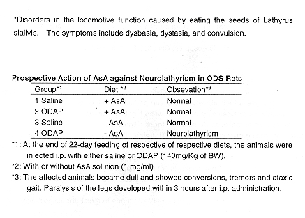
not enter the brain because of the BBB. AsA-deficient rats abdominally administered this amino acid showed lathyrism*, or neural symptoms, whereas the controls do not show any change. Rats with the neural symptoms stop their locomotion and tremor, paralysis of legs, etc. are observed. These symptoms are presumed to be caused by the entrance of the neuro-stimulating amino acid to the brain because of damage or change in the function of the BBB induced by AsA deficiency. Such symptoms are actually found in people in poor nutritional conditions in developing countries after they eat beans grown for food and decoration and containing this substance. The causal substance of lathyrism contained in the beans, 3-N-oxyalyl-1,2,3-diaminopropane (ODAP), has a direct effect on neural receptors, resulting in the liberation of zinc and the formation a strong chelate with this zinc, causing zinc deficiency. Zinc is liberated at glutamin-dependent synapses upon neural stimulation and is believed to regulate the level of neurotransmission by glutamic acid. Patients with lathyrism show decrease in zinc levels in the blood. It is likely that excessive neurotransmission occurs in a zinc- deficient brain, causing functional disorders.

In the above, I have introduced part of our study showing that AsA deficiency causes not only disorders in the neurotransmission system but also morphological changes in neurons, and that the same deficiency causes cerebrovascular damage which induces a change in the BBB function, resulting in changes in the permeability to substances entering the brain. 
The homeostasis of the brain is maintained better than any other organ. However, the changes in brain functions caused by AsA deficiency seem to be an example showing that changes of the amount of dietary nutrients, especially essential trace nutrients which cannot be synthesized in the body, may affect brain functions through changes in the state of the BBB even in the brain with a fully-developed BBB.
The increase of the number of patients with brain disease such as senile dementia caused by abnormal aging of the brain is also a major problem facing Japan in the 21st century. In the future, it is necessary to further study the effects of environmental factors on the BBB, especially of nutrients which are directly connected with our daily diet, in terms of brain functions. The analysis of the complex mechanism with multiple effects is awaited, such as the effects on the brain and other organs when digestion and absorption are taken into consideration, and the interaction among various substances when permeating into the brain. 


Determination of Efficacy of Tanning Prevention Products
F. Hoffmann-La Roche Ltd, Vitamin and Fine Chemicals
Cosmetic Research, CH-8600 Duebendorf, Switzerland 


Introduction
       Skin lightening products and fairness creams promise beside the control of pigmentation the protection from UV-radiation induced tanning. The normal sunscreens claim and express their efficacy as the sun protection factor (SPF), telling the consumer, how much longer he can stay in the sun without getting sunburned, if the product has been evenly applied to the skin. In contrast, for skin lightening products no quantitative efficacy claim for tanning prevention is made. Therefore, the consumer has no indication to what degree he is protected from tanning and he has no opportunity to compare commercial fairness creams based on their efficacy of tanning protection.
       The protection from tanning is not only depending on the concentration of UV-filters added, but depends very much on the combination of UVA- and UVB-filters and on the vehicle. The UV-action spectra responsible for tanning is known, as well as the irradiation spectra of the sun. The combination of UV-filters has to match the combination of these two spectra for optimum protection.
       This tanning prevention factor is a quantitative mean to compare the efficacy of different tanning prevention products, as the SPF (Sun Protection Factor) allows to compare UVB-protection of different products. It dose not describe the fairness activity of skin lightening cream. However, skin lightening without tanning prevention is pointless. Even if people would not leave the house or remain always in the shade tanning will occur, since UVA-radiation does penetrate window glass to a large extend (80%), and the reflected or scattered part will reach the skin even in the shade. Therefore, all fairness products should contain UV-filters for tanning prevention and should claim their degree of protection.

Method
       The method developed to quantify tanning prevention by sunscreens is build on the method for SPF determinations, the efficacy claim for UVB-protection. Unprotected skin is irradiated to determine the minimal dose necessary to provoke a tanning on human skin. The tanning protection product is applied in a concentration of 2 mg/cm2 to an other area of the human volunteers back, allowed to dry for 15 minutes, and is irradiated in the same way, using 6 dosed differing by 30% of UVA. The measurements are conducted under GCP (Good Clinical Practice) and according to the roles set by the WHO for clinical testing on humans. A case report form (CRF) is used for each volunteer.

Irradiation conditions
       A metal-halogen lamp of 400 W (Philips HB853) is used, with a WG320 filter to remove all UVC radiation. The distance between lamp and skin is adjusted to have an irradiation intensity of about 180 mJ/ cm2. Depending on the uniformity of the volunteers back, 6 test fields are marked. The exact irradiation intensity on every field is measured and used to calculate the irradiation time of the single spots. A template is placed on the back of the volunteer, which has 6 small flaps in every test filed, covering the irradiation spots. Six spots of 1 cm2 each, are irradiated with UVA-doses increasing 30% from spot to spot.

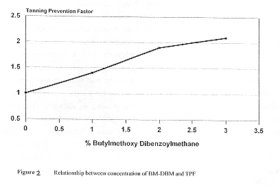
Calculation of Tanning Prevention Factor (TPF)
The minimal dose to induce a tanning on the protected side of the skin divided by the minimal tanning dose for unprotected skin area leads to the tanning prevention factor.


Discussion
       The different color tones of human skin are a combination of melanin, hemoglobin, carotenoids and the condition of the stratum corneum. The red color is due to a blood pigment called oxygenated hemoglobin, the yellow is due to the pigments called carotenoids, the blue color is due to the "reduced" hemoglobin in the veins, and the major brown color is due to the presence of the pigment melanin, which is produced in the melanocytes. In the basal layer of the epidermis there are melanocytes and basal keratinocytes in a ratio of about 1:4 to 1:10. One melanocyte will provide melanin pigment to about 36 keratinocytes. The association of melanocyte with these keratinocytes has been called the epidermal melanin unit.
       Melanin produced in the makanocyte is transferred to the keratinocytes in packets called malanosomes. The malanosomes are transferred to the keratinocytes by dendritic projections, a process very similar to the process of injection with a hypodermic needle. Negroid malanosomes are larger than those of Caucasians and this may contribute to their differences in skin color. Furthermore, melanocytes in dark skinned people are more active, producing more melanin, and in the epidermis of dark skinned people melanin is not reduced by enzymes.
       The initial steps of the melanogenesis are under control of the enzyme tyrosinase, which oxidizes tyrosine to 3,4-dihydroxphenylalanine (DOPA) and then to DOPA-quinone. DOPA-quinone is concerted by a series of complex reactions involving cyclization and oxidative polymerization, which finally results in the formation of eumelanin. If sulphydryl residues are available from cystein or glutathione cysteinyl-DOPA is formed and quickly oxidized into benzothiazines and thence into pheomelanins.
       Eumelanin is the brown-back melanin seen in brown skin and black hair. Pheomelanin is the red-yellow seen in red hair. Pigmentation in man is regulated mainly by two mechanism. Constitutive pigmentation is the level of pigmentation based on a persons genetic predisposition, in part of the body that are not normally exposed to UV-radiation or any other stimulatory influence. Facultative skin pigmentation is the tanning induced by either UV-radiation, a major factor, or hormones.
       Tanning by UV-radiation (sun light) involves two distinct reactions. The first, or immediate reaction, which occurs within a few minutes of exposure to UVA and which will last for up to 2 hours, is characterized by the photo-oxidation of preformed melanin. The delayed action which occurs in response to both UVA and UVB is much slower in onset and is only apparent after 2 to 24 hours of exposure. This delayed response involves an increase in the number of active melanocytes, enhanced melamosome production and an increase in melanogenesis. There is also an increase in keratinocyte proliferation and the transfer of melanosomes from melanocytes into these cells.
       Figure 1 shows the solar UV-spectra and the different action spectra. The solar spectra was measured at a clear summer day, in July at noon, at latitude of 55°north. The action spectra was measured by Parrish and Rosen (1990). Such action spectra are highly skin type dependent. Even if these curves are only approximations, they nevertheless give a good idea at which wavelength an UV-protector has to be active to prevent a physiological stimulation or a certain damage.

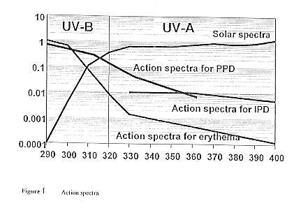
       Tanning prevention is different from general sun protection. Normal sun protection is assessed on its efficacy to prevent an erythema.  As the action spectra for tanning is different to the action spectra for erythema, so is the optimum filter combination different for the two purposes. It has to be taken into account, that of the total UV-radiation reaching the earth surface only about 20% (21.1 W/m2) is UVB-radiation and 80% (85.7 W/m2) is UVA-radiation. The protection from UVB-radiation is expressed in the SPF. Various methods are available and also comparable, since all the products use defined solar simulated irradiation spectra and differ mainly in the statistical treatment of the results, the number of volunteers and the size of the application fields. Similar to the SPF we developed a method to quantitatively assess the protection from UVA-radiation and therewith the efficacy of tanning prevention on skin.
       The UVA-spectra used for irradiation originates from a medium pressure metalhalogen lamp of 400 W. The UVB-radiation is filtered-off by a filter. The minimal tanning dose for skin type V (Indian skin) is in the order of 20-30 J/ cm2. This dose is usually obtained within 10 to 20 minutes. The lamp intensity is about 5 times higher than normal sunlight.

Conclusion
       Figure 2 shows the linear relationship between UV-filter concentration (BM-DBM) and the TPF. This is the result obtained with a typical O/W emulsion. Other formulations may lead to different absolute values. In addition to BM-DBM the emulsion contains UVB-filters (octylmethoxycinnamate and octocrylene). A TPF of 3 dose approx. correspond to a 4-5 hours direct sun exposure in Bangkok. This method allows quantitative comparison of different tanning prevention products and to quantitatively assess the tanning prevention properties of sunscreen products.


Oral Contraception and Nutrition No.4
(from Nutritional Concerns of Women)

Impairment of Folic Acid Status
       There is evidence that OCs interfere with and impair the body's metabolism of folic acid, or folate, and vitaminn B12. However, the following two sections will show that several investigators have reported results that negate these findings. This discrepancy, independent of biochemical indicators used, has created some confusion and controversy.
       Clinical and biochemical indicators frequently examined to assess the folate nutritional status have been the following: (1) the incidence of megaloblastic anemia and cervical dysplasia, (2) serum and erythrocyte folate concentrations, (3) the urinary formiminoglutamic acid (FIGLU) excretion, and (4) the intestinal absorption of polygultamates. Numerous cases of megaloblastic anemia attributed to OCs have been reported. In some of these cases there were associated contributory factors, such as a mild malabsorption syndrome or dietary folate deficiency, and it is not clear whether megaloblastic anemia would have occurred in the absence of these factors. The literature pertinent to the association between OC use and folic acid status is divergent. Some reports that have demonstrated that women using OCs have a lower serum level of folate than nonusers have been contradicted by others showing no statistical difference. The subject is also considered controversial when the erythrocyte level (a better biochemical marker than serum level and more representative of issue metabolism) is used to assess nutritional status. A number of investigators have reported that the mean red cell level of folate in groups of women taking OCs, as assessed by microbiological techniques, was significantly lower than that of control groups. Other studies have failed to show any statistical differences.
       Although conflicting data still exist as to the effects of OCs on folic acid status, the majority of reports tend to support the observation that there is a parallel lowering of the folic acid concentration in erythrocytes and an increase in Urinary FIGLU excretion simultaneously with the reduction in serum folate levels. FIGLU is an intermediary product of the metabolism of histidine that requires the reduced form of folic acid to be further metabolized. Shojania found that women using OCs excreted significantly more FIGLU in the urine after a histidine load than controls and that the levels decreased to normal within 2 to 4 months after their use was stopped. It is not clear whether this increase is due to folate deficiency induced by OCs or to a physiologic effect that mimics early pregnancy. A higher level of FIGLU is also excreted during pregnancy.
The mechanism of the impairment of folic acid metabolism by OCs, both in experimental animals and in humans, remains unclear. The difference in the intestinal absorption between the usual dietary folate and folic acid in therapeutic vitamin preparations, in OC users, has been underlined in several medical reports. Polyglutamates, the major dietary source of folic acid, must be enzymatically deconjugated in the small in the small intestine before absorption takes place. Streiff has described cases of women on OCs showing a defect in utilization or absorption between polyglutamates and monoglutamate.
       Initially, the inhibition of the activity of folate conjugase by OCs and the consequent malabsorption of folate polyglutamates seemed to have provided a statisfying explanation for the imparment of folic acid metabolism. However, a subsequent report failed to show any malabsorption of folate polyglutamates in OC users or any inhibitory effect of these compounds on folate conjugase. The hormonal alterations found during pregnancy do not selectively change polyglutamate absorption, suggesting that oral synthetic estrogens and progestogens may act in a different manner than do the naturally occurring hormones, at least as far as intestinal absorption of folic acid is concerned. Another mechanism has been proposed for the impairment of folic acid metabolism in association with the use of OCs. The increased excretion of folates in the urine, which is also reported in pregnant women, may in part explain the lower levels of folic acid in the serum and erythrocytes of some OC users.

Impairment of Vitamin B12 Status
       OC users have been reported to exhibit significantly reduced levels of serum vitamin B12 whether the level is determined by microbiologic or radioisotope assays. It is difficult to explain the mechanism behind this phenomenon. Serum levels of vitamin B12 may be lowered, to subnormal values in some cases, but this finding is not necessarily associated with evidence of tissue depletion (sign of a true deficiency). However, the clinician may suggest that the patient temporarily stop taking "the pill" to see if the serum level of vitamin B12 is decreased. A Schilling test (a radioactive absorption assay) is usually performed to exclude the possibility that the problem is caused by vitamin B12 malabsorption. Results from our current logitudinal (six menstrual cycles) study on young women taking TriphasilR in regard to vitamin B12 and other nutrients are summarized in Table. Mean serum vitamin B12 level was reduced by 26% but the difference was not significant (due to high variance of data), despite the fact that the blood sample was withdrawn at the same period of menstrual cycle for all subjects.
       Besides malabsorption problems, proposed explanations for the serum vitamin B12 reduction have included an increased renal excretion and an impaired production of vitamin binders. Hjelt er al. measured vitamin B12 absorption and excretion by means of a sensitive whole-body counting technique. Since none of the OC users suffered from dietary insufficiency, the findings of normal absorption and excretion indicated that the vitamin B12 stores were normal. This is in accordance with others who have found a normal Schilling test and no changes in erythrocyte levels and urinary excretion of methylmalonic acid (another biochemical marker for vitamin B12 status).
       The low serum vitamin B12 level is most likely associated with defective serum vitamin B12 binders. In fact, Ocs can inhibit the production of transcobalamin I (TC-I), a glycoprotein synthesized by leukocytes. This binder, 70 to 100% saturated, is not essential for vitamin B12 transport to the tissues and is more concerned with its transport in plasma. Total serum level of TC-I has been found to be reduced or not significantly changed in OC users. Since about 90% if vitamin B12 is bound to TC-I, a low level could contribute to the low serum vitamin B12 level. TranscobalaminUis a beta globuline and is 90 to 95% unsaturated, but it plays the major role in vitamin B12 transport to the tissues. Larsson-Cohn suggested that OC treatment increase the tissue avidity for vitamin B12 resulting in a redistribution of the vitamin within the different tissue compartments. This is unlikely to occur according to Shojania and Wylie, because TC-U is unaffected by OCs. 


AMD and LuteinA
(from Nutritional and Environmental Influences on the Eye) 
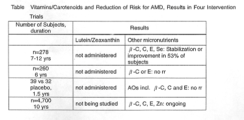

INTERVENTION TRIALS
       Unfortunately, there are no results of human intervention trials to date using lutein and/or zeaxanthin since these are of limited availability for human consumption. Also neither carotenoid is being used in the ongoing major clinical study called Age-Related Eye Disease Study (AREDS) initiated by the National Eye Institute. Four intervention trials using related micronutrients and studying opthalmologic parameters tabulated in Table.
       The only intervention data with zeaxanthin so far available were obtained in animals. Four three months quails were fed carotenoid free diets or diets supplemented with 5 mg/kg 3R,3'R-zeaxanthin. The animals were then exposed to intermittent white light (3200 lux) for 28 hours in order to induce general photic damage to the retina. After 14 hours in the dark, the eyes were excised for the determination by HPLC or zeaxanthin in the retina and the measurement of the extent of apoptosis, which was confirmed by TUNEL stain, The number of light-induced apotoses of rod and cone photoreceptor cells was drastically reduced in treated vs untreated animals. Furthermore, the retinae containing more zeaxanthin as assessed by HPLC seemed to be better protected than those with less. This is the first direct preclinical demonstration of the efficacy of zeaxanthin for the prevention of one important consequence of light-induced retinal damage. It complements older data showing loss of macular pigment and more drusen and other indicators of increased photic damage in monkeys raised on a carotenoid-depleted diet for 5 years.